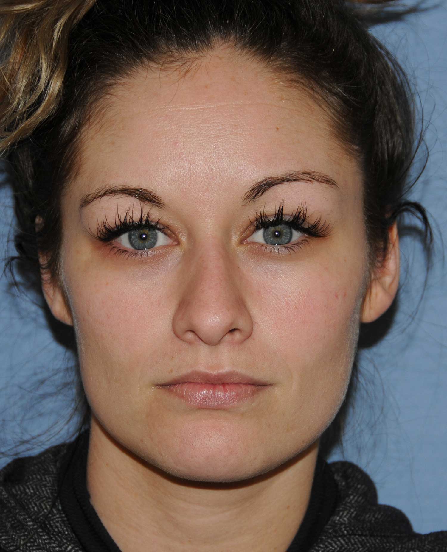
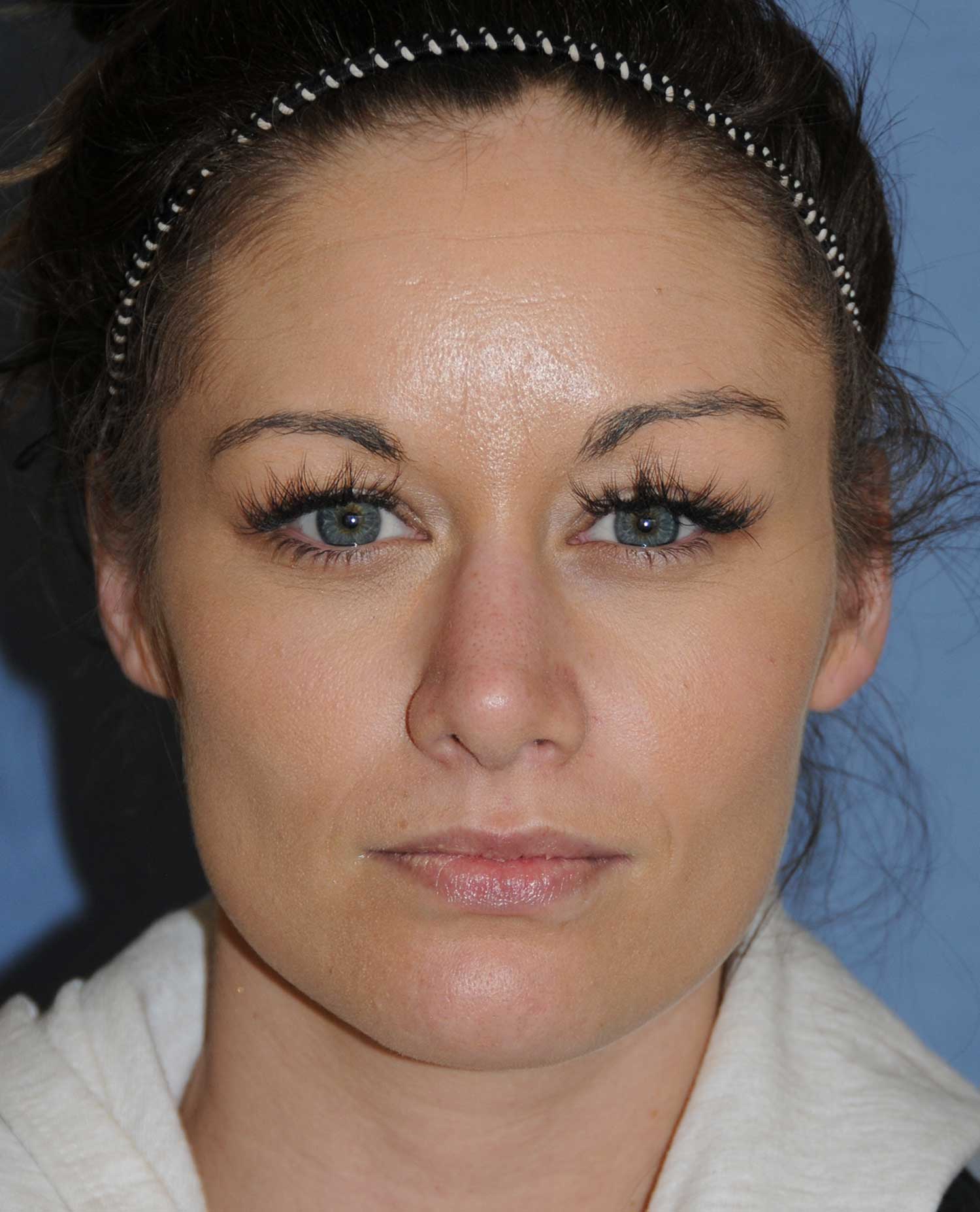
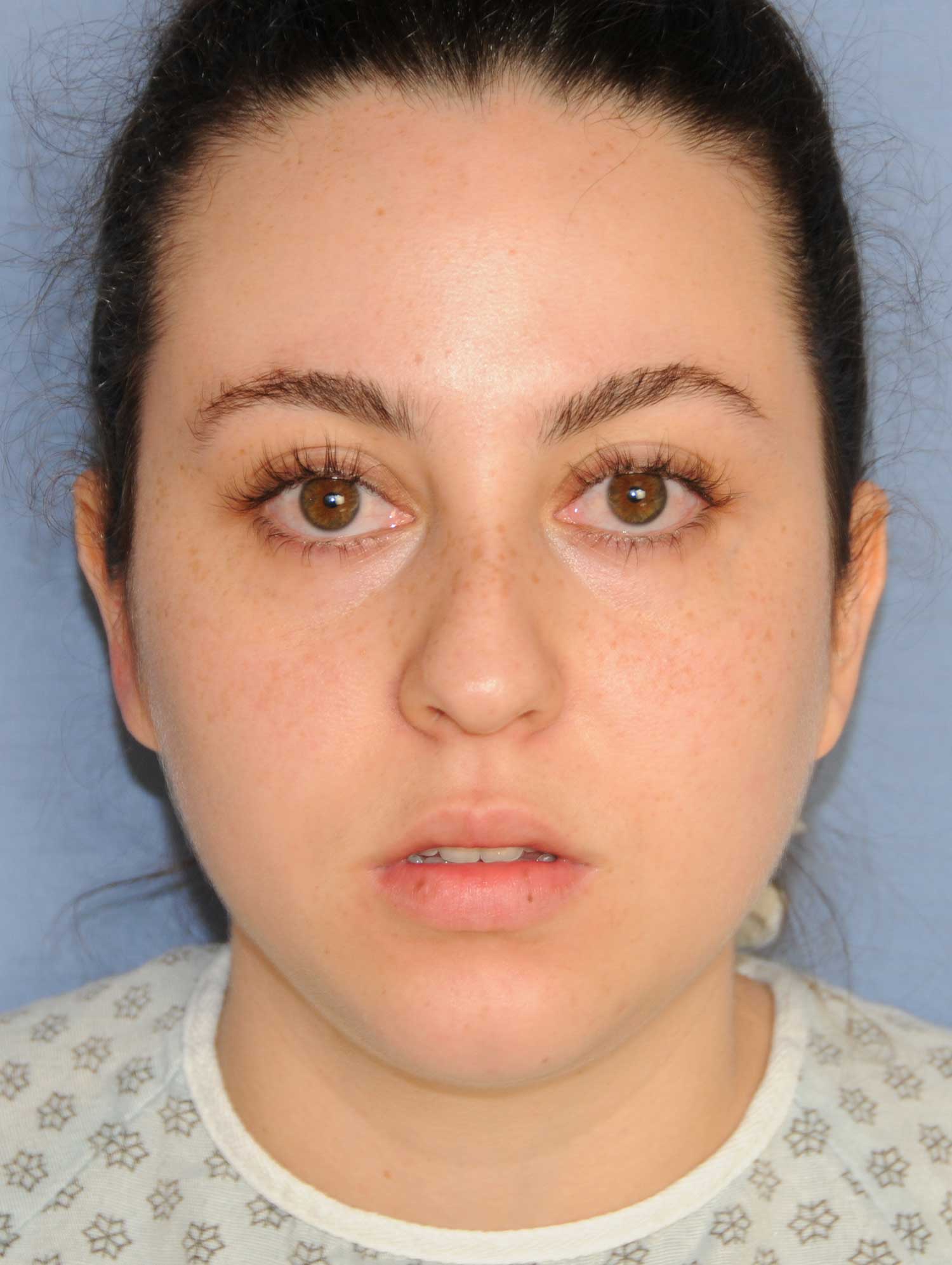
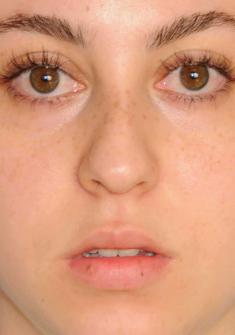
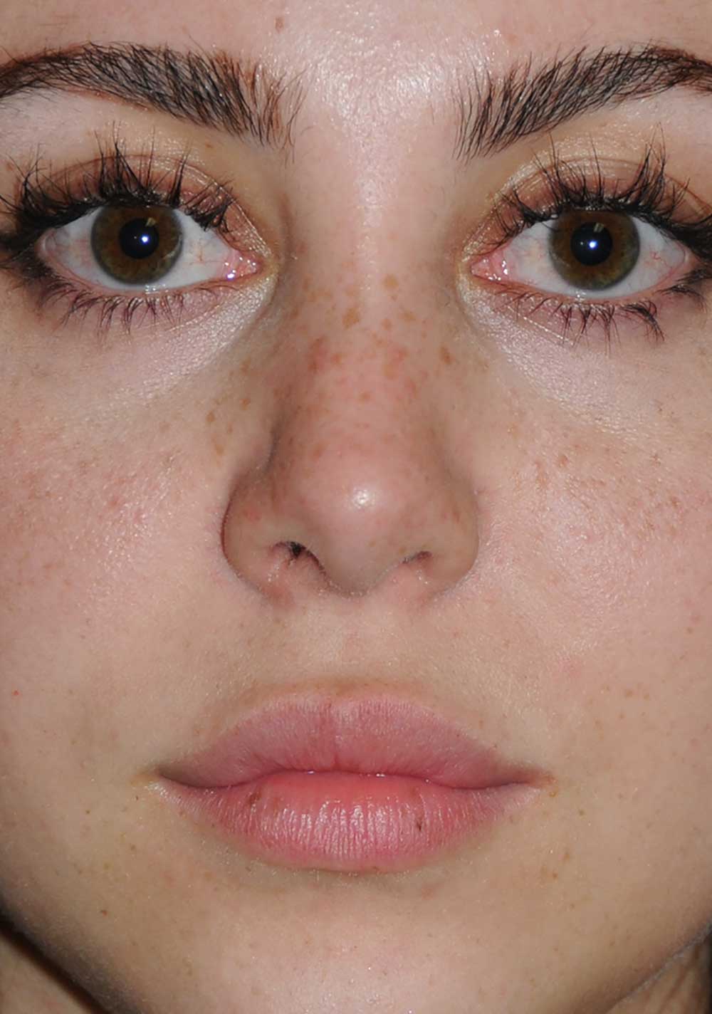
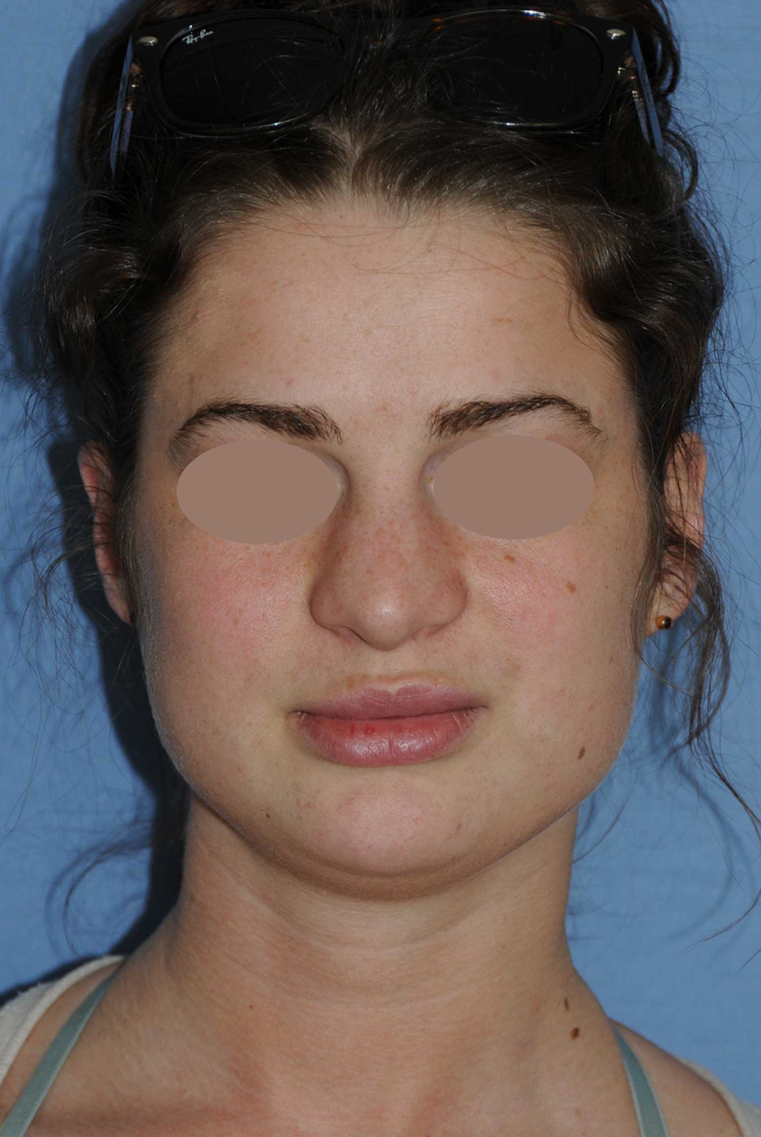
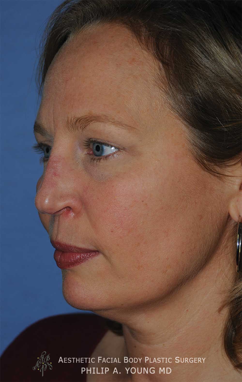
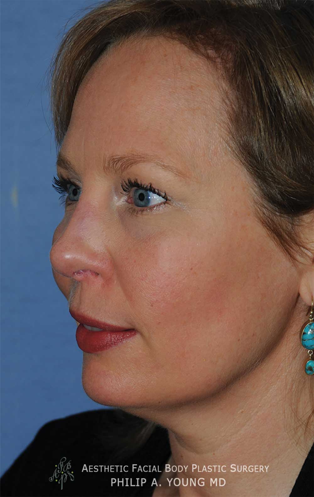
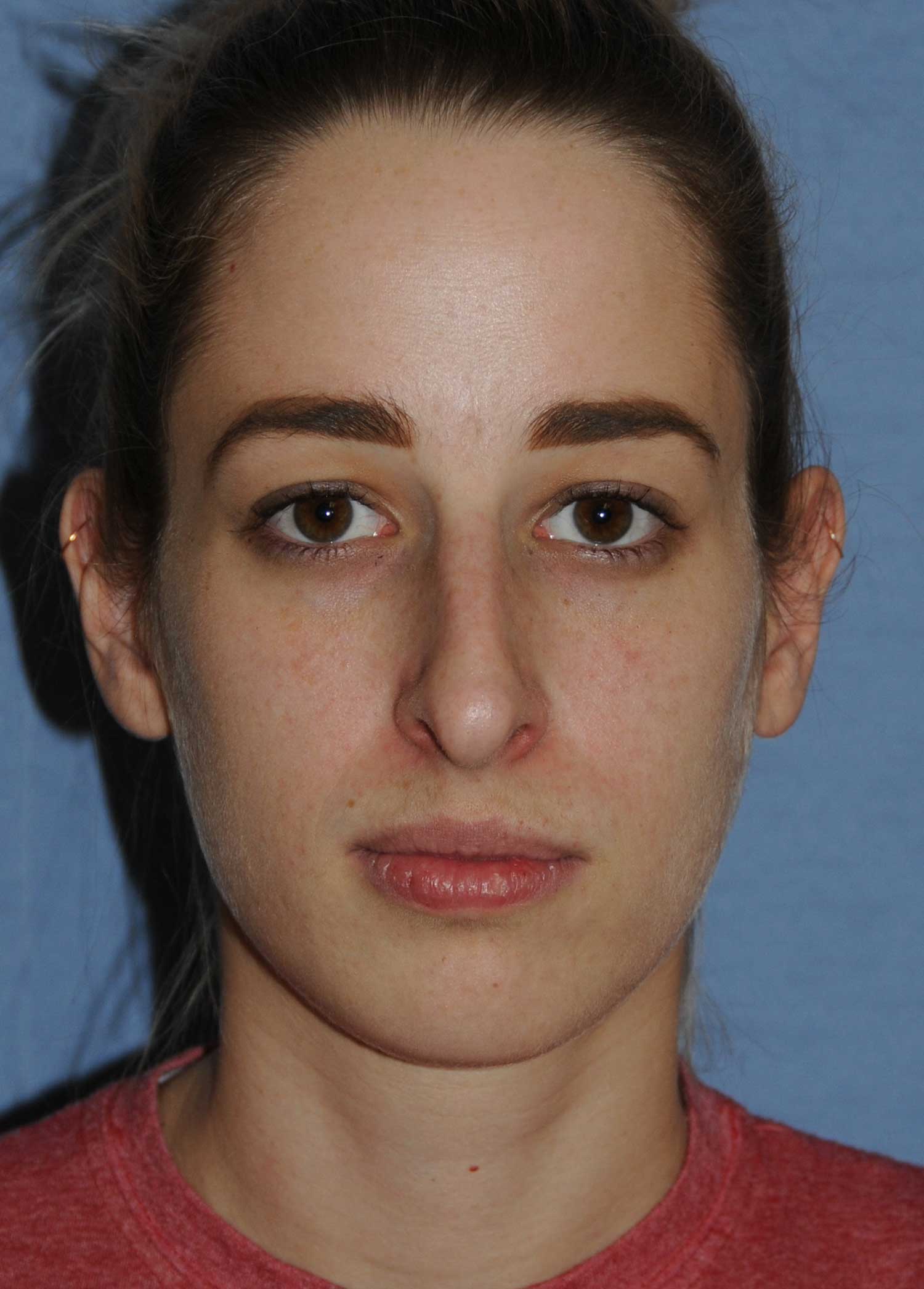
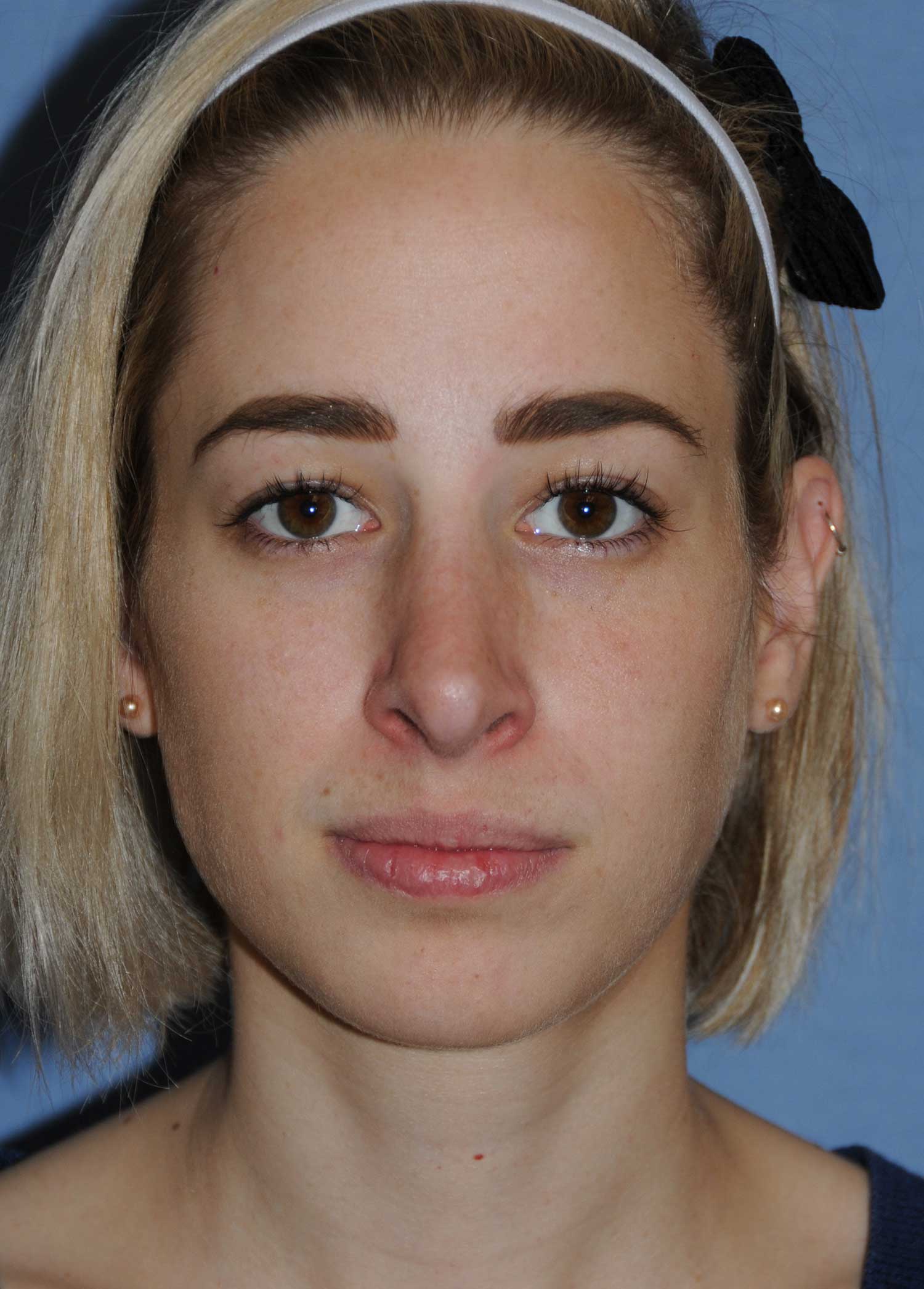
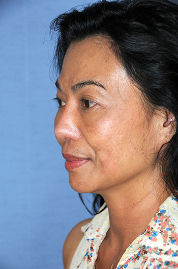
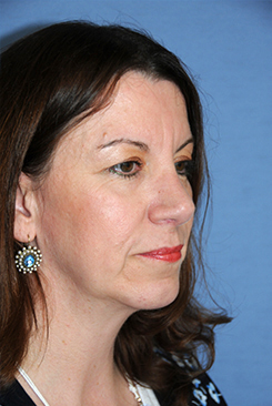
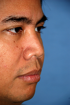
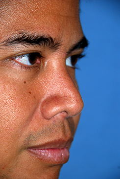
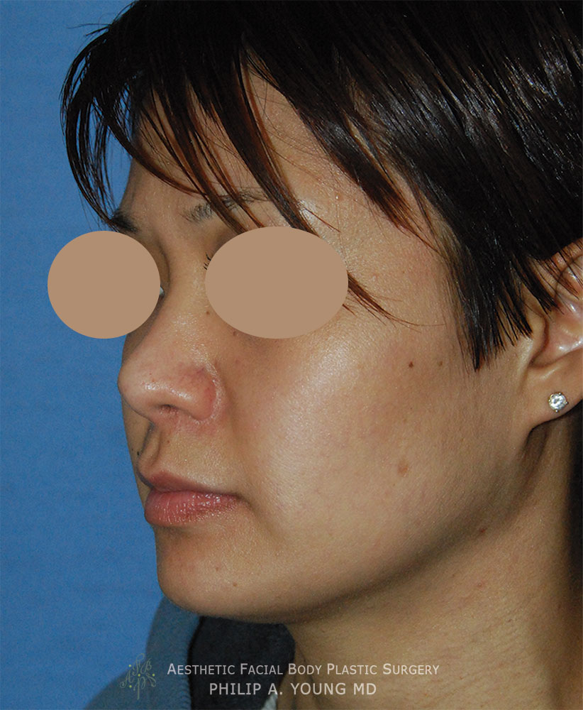
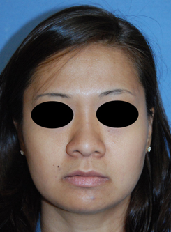
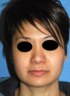
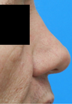
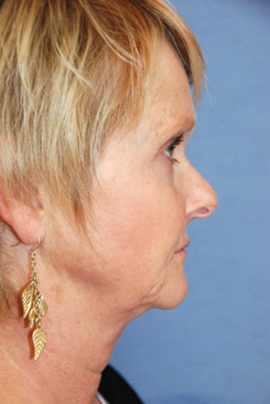
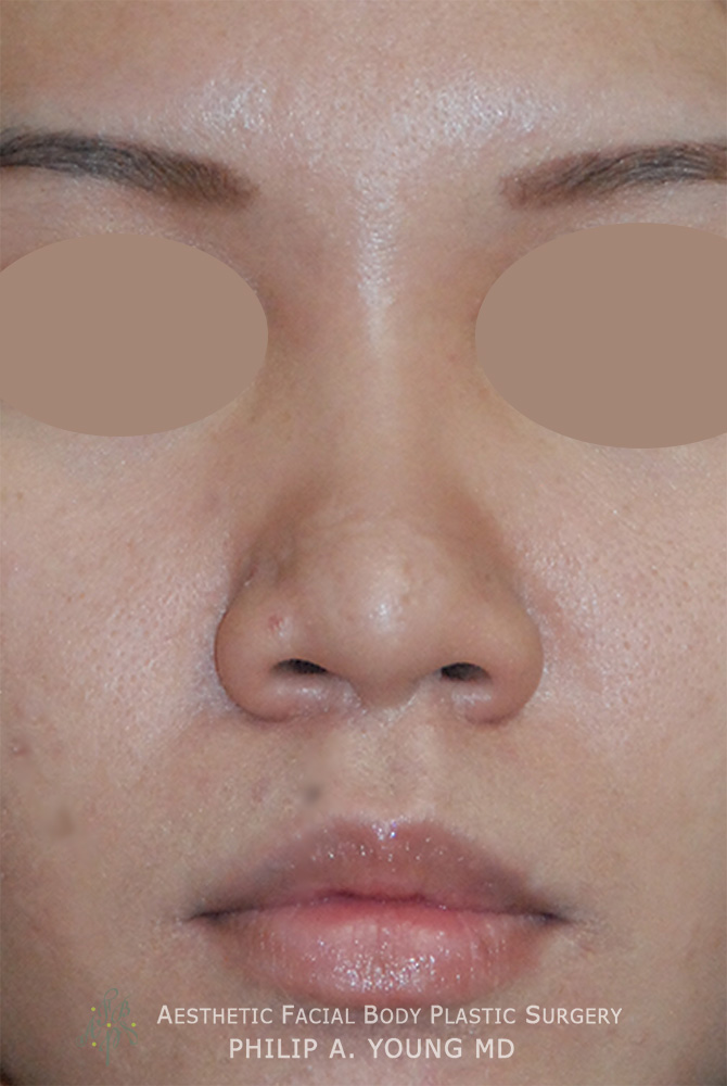
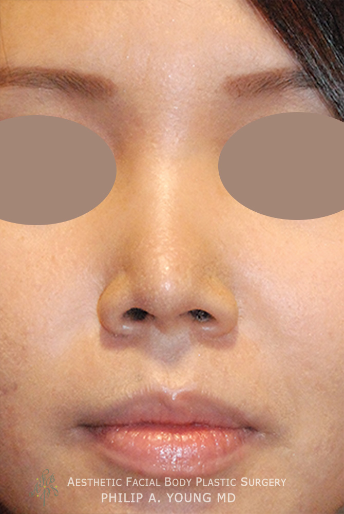
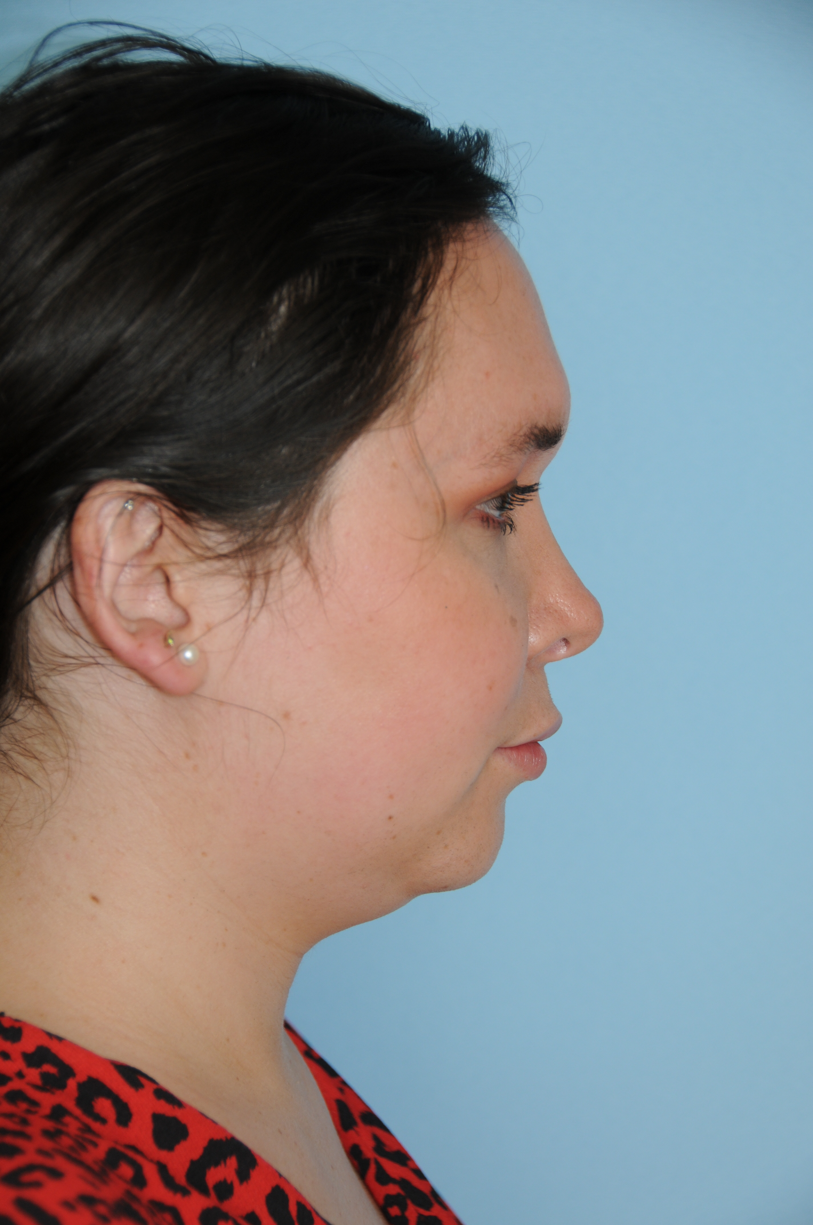
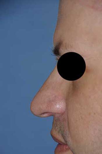
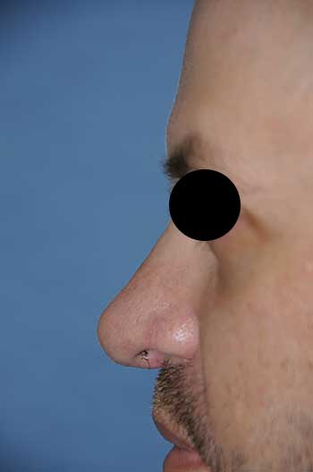
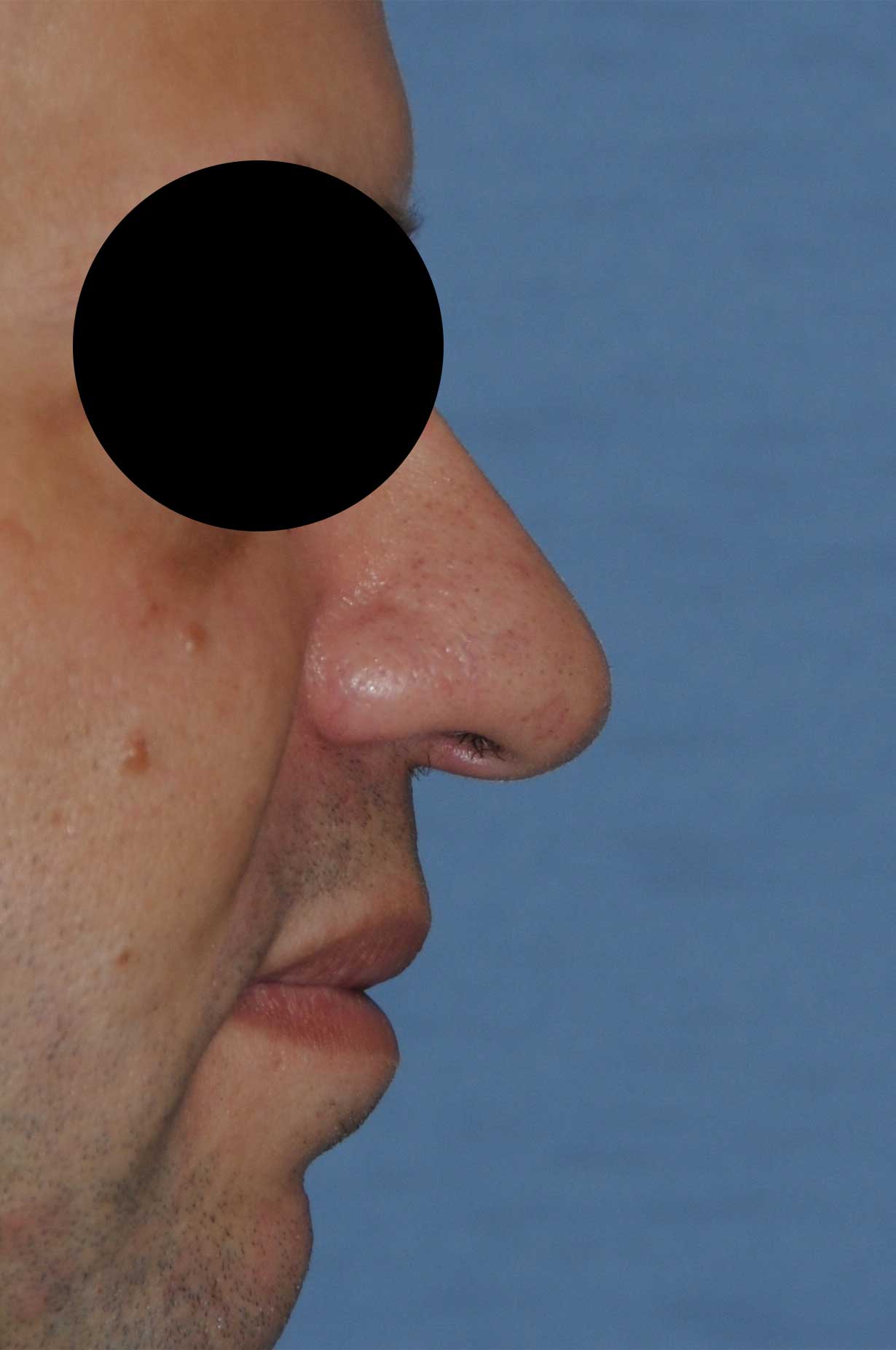
This is Seattle Rhinoplasty Surgeon Dr. Philip Young and Aesthetic Facial Plastic Surgery’s Rhinoplasty page dedicated to the many terms used in Rhinoplasty in a dictionary type of approach. We give a brief description | definition and then we tell you the most relevant significance of this area in relation to the Rhinoplasty procedure:
Accessory Cartilage: These are the cartilage tissue that is connected by fibrous bands that connect to the lower lateral cartilages and the pyriform aperture (bony ring around the nose made of maxilla, nasal bones, frontal bone (nasal and maxillary process). They are important in that they give this area some rigidity and helps with the airway.
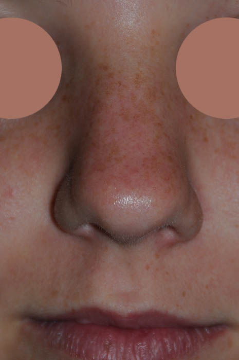
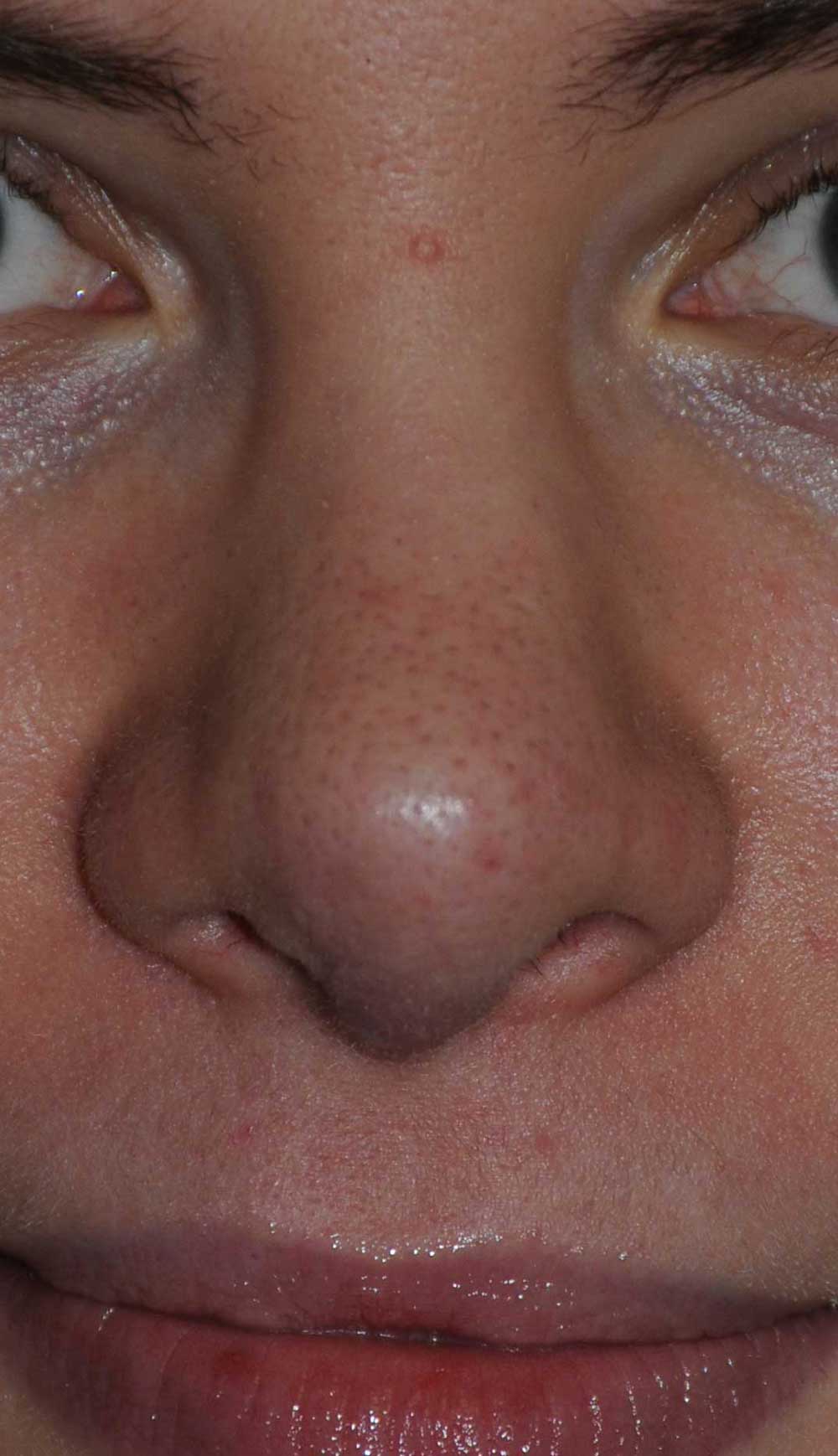
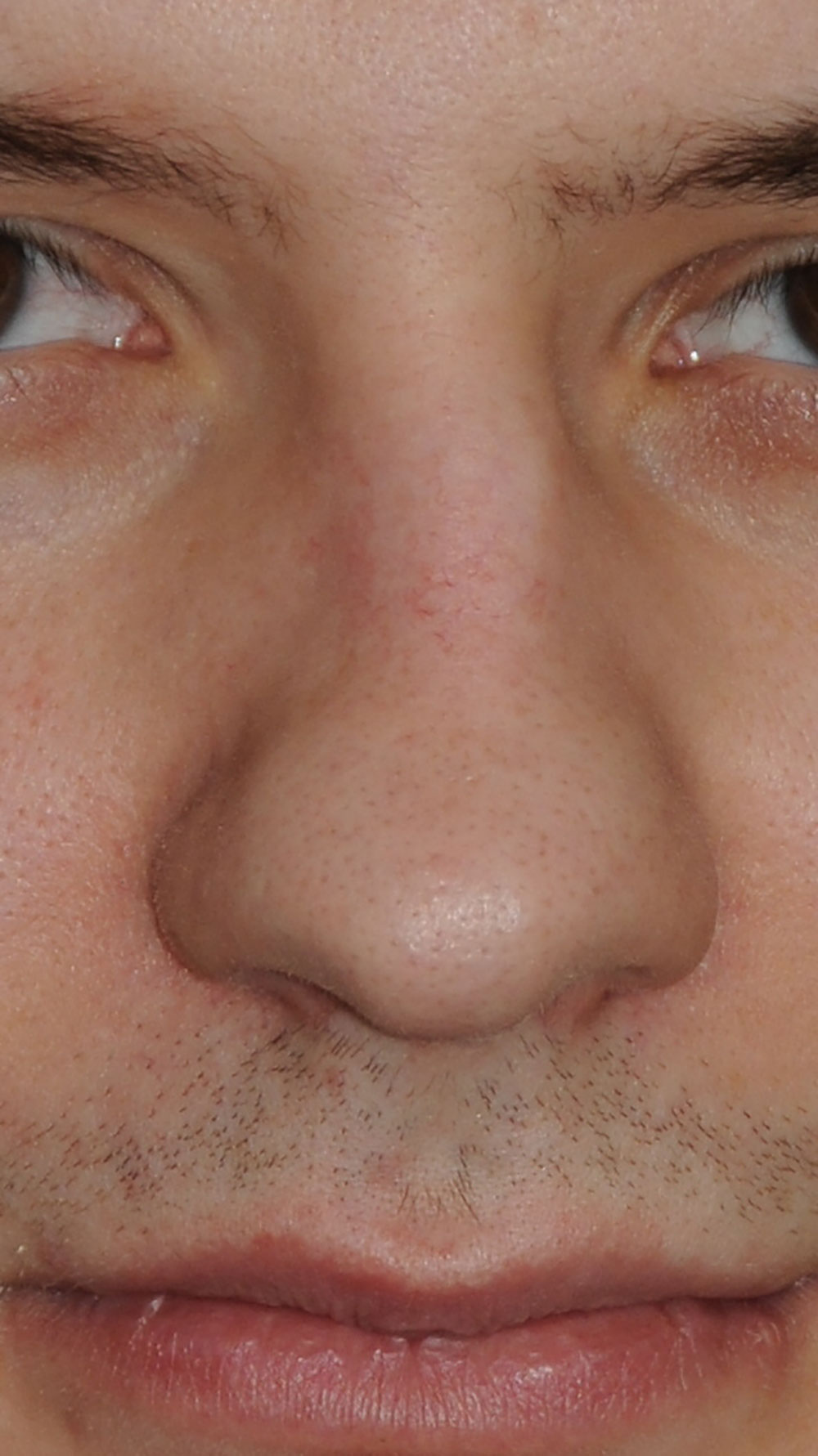
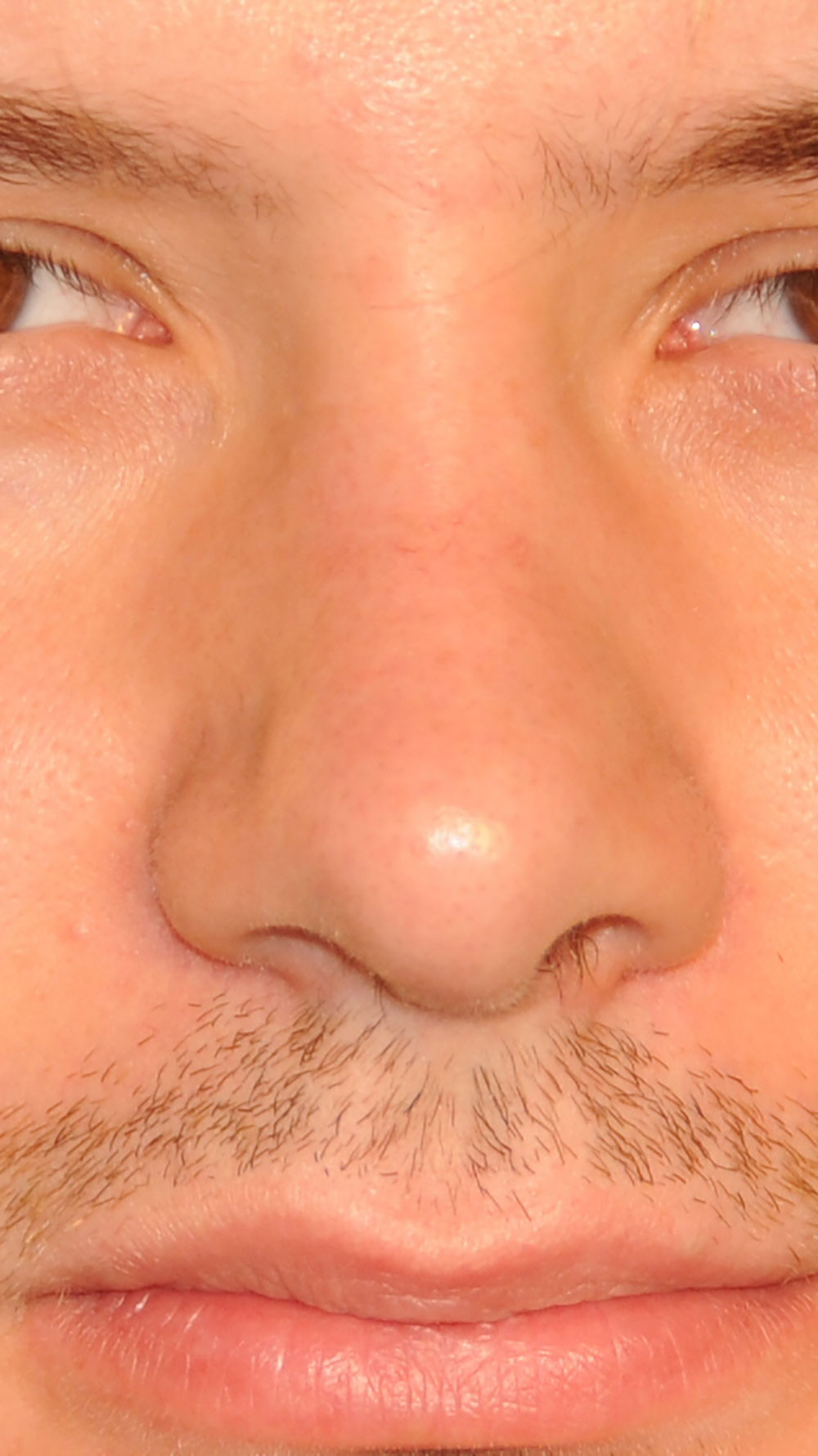
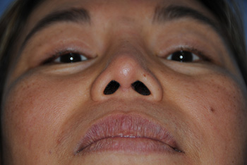
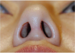
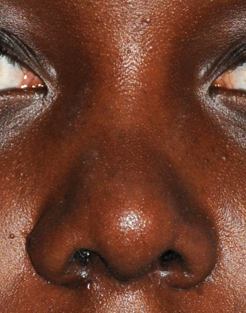
Alae: This is the fibrofatty tissue that envelopes the alae. It is made without cartilage and is consistenting of only fatty tissue with collagen, elastin, skin, some nasal muscles and the covering of the muscles (fascia). You can make incisions on the outside or within the nostrils to make the base of the nose narrower and alter the shape of this area.
Alar Base: is defined most commonly as the area that is occupied by both Alae and the base of the nose at this location. The thought is that the based is mostly preferred to be lined up with the medial canthi (inner corners of the eyes). Alar Wier Excisions on the outside can narrow the base along with resection of the nostril rim inside the nostrils.
Alar Groove: is the linear depression between the ala and the nasal sidewall where the upper cartilages reside inside.
Alar rim graft: is a very common graft that is placed along the inferior edge of the alae in order to support this structure. These grafts tend to bow out the rim and are used in cases where the nostrils are collapsing.
Anatomic Dome: The most anterior projecting part of the tip that occurs at the junction where the lateral crura and intermediate crura connect superiorly.
Anterior Septal Angle: This is the angle that is formed by the dorsal and caudal struts of the septum. This angle is very important for determining the position of the tip. Altering this can have dramatic impact on the tip position, angle of the lower nose of side view, nasal length, tip projection, tip rotation, etc.
Binder Syndrome: Is a developmental disorder primarly affecting the nose and the maxillary bone. Causes are unclear but research has centered around genetics and Vitamin D deficiency. Can include absense of the anterior nasal spine; under developed nasal septum, premaxilla or the anterior part of the maxillary bone that makes up the palate; anomalies of the muscles of the upper lip, nasal floor and cervical spine. An under developed upper jaw, class 3 occlusion or underbite is seen with this condition as well. Skeletal Distraction at age 8 is sometimes done along with orthognathic surgery and other plastic surgery procedures.
Bony Vault: Is the bone around the nose that comprises of the maxilla that is the lateral and inferior edge of bone, the nasal bones which is superior and the nasal process of the frontal bone.
“My goal was to find a method to bring back a person’s natural youthfulness without the operated, unnatural look and that is why I have my patients bring in photos of them when they were age 5 to 30. My YoungVitalizer helps restore natural and youthful contours they haven’t seen in years.”
—Dr. Philip Young, Seattle Facial Plastic Surgeon

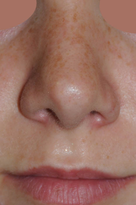
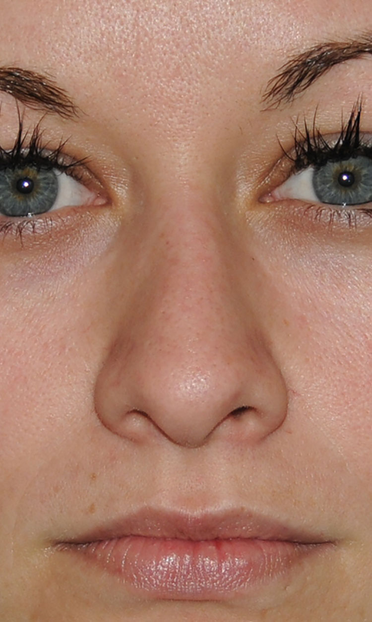
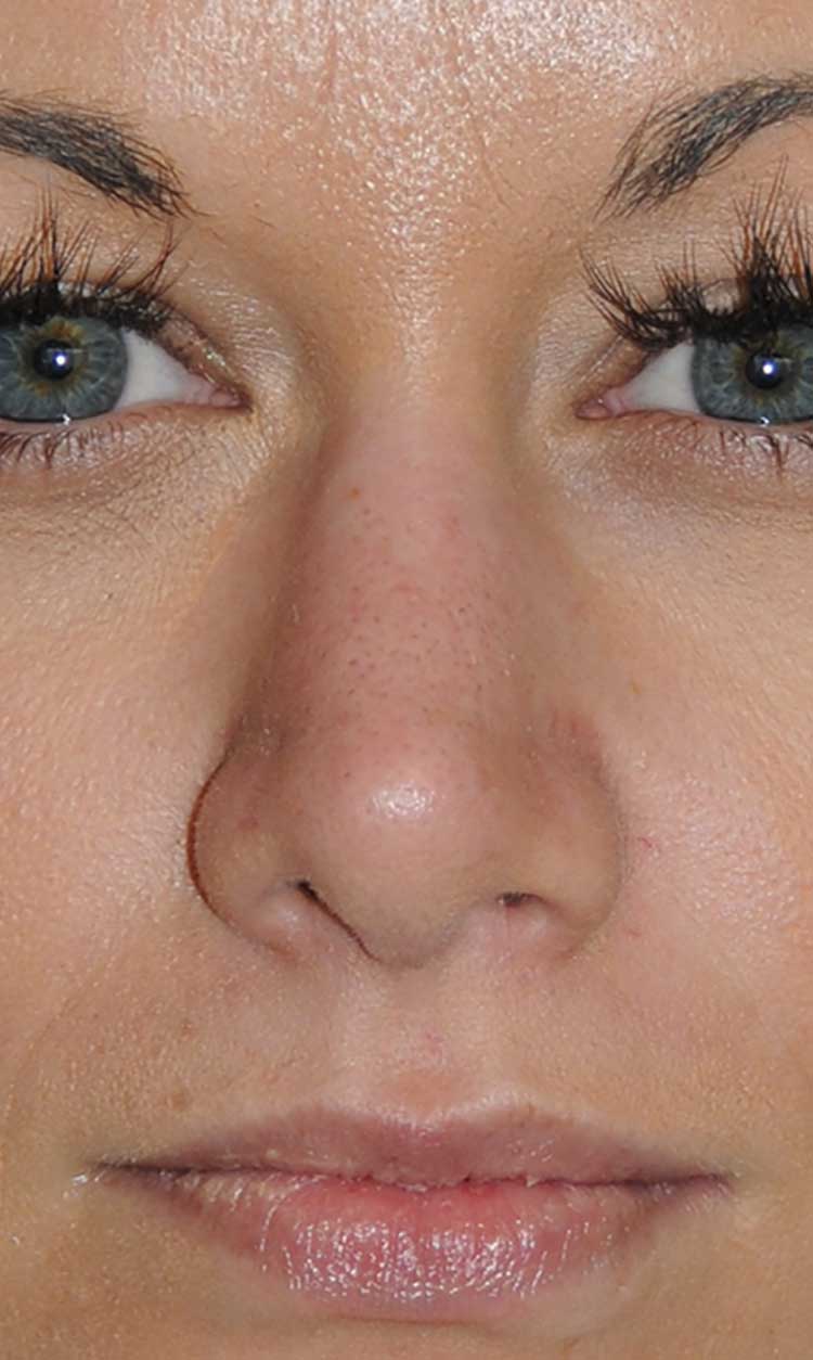
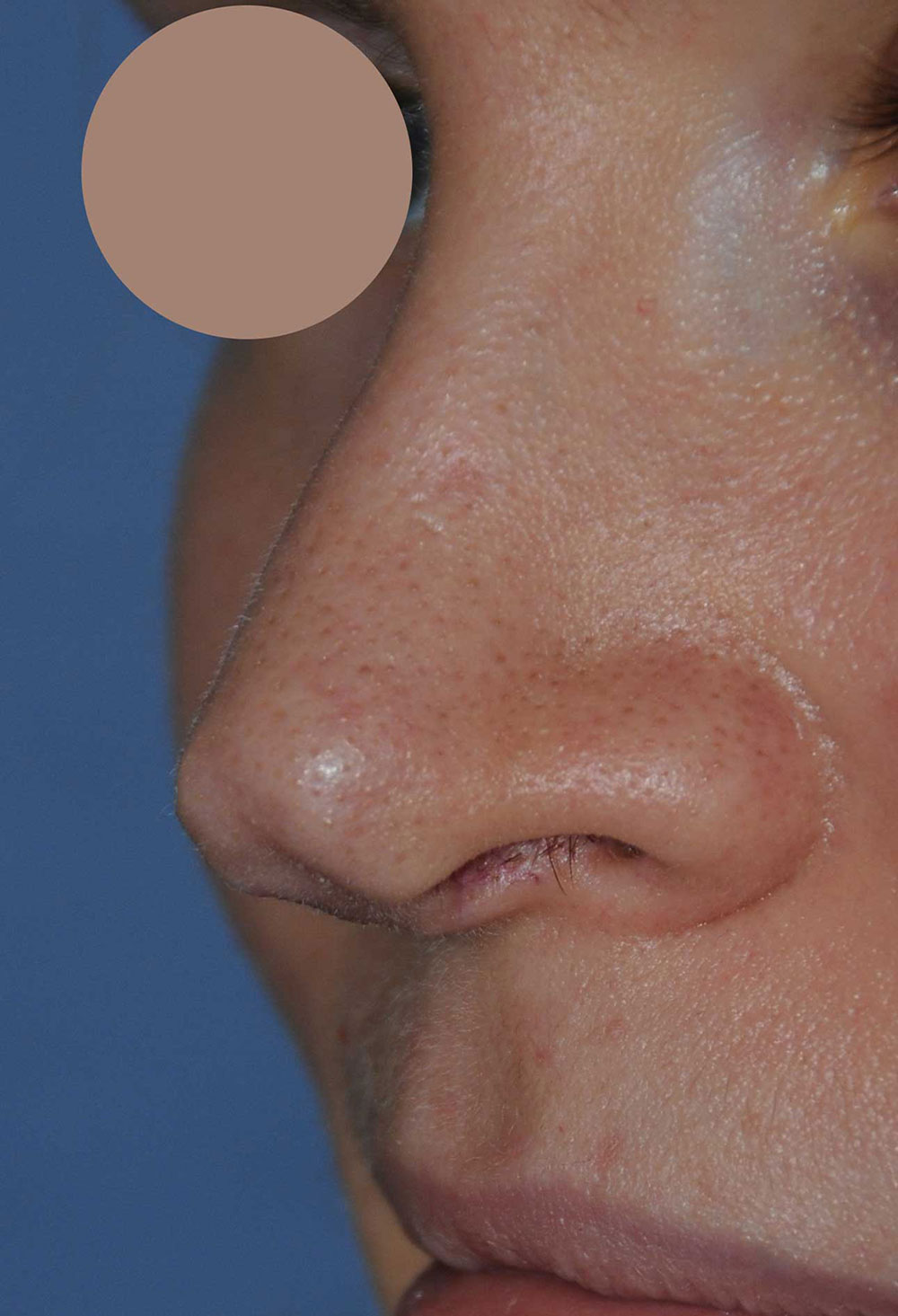
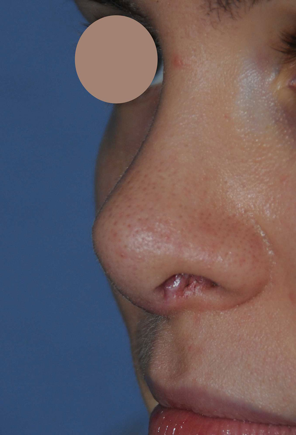
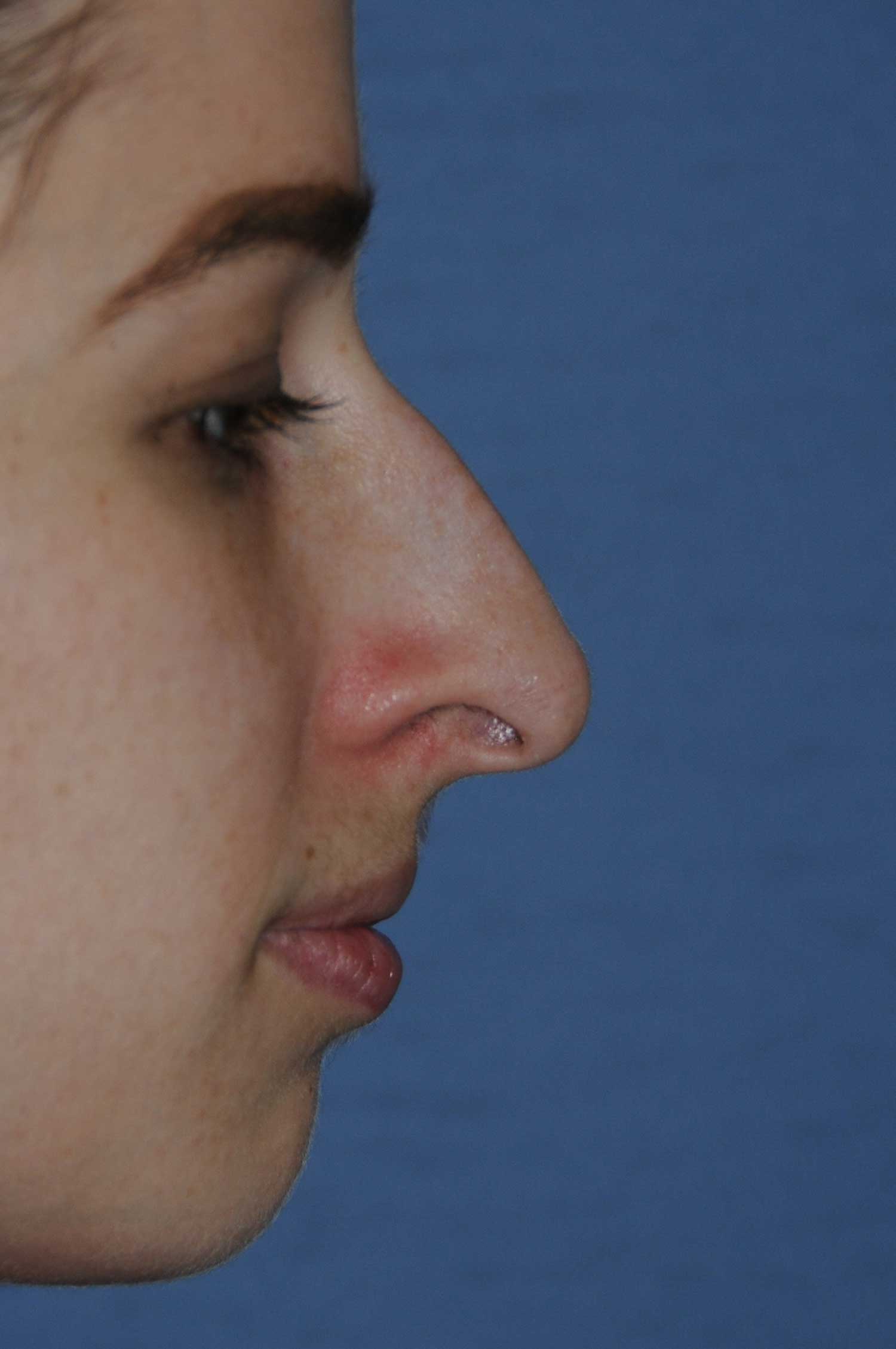
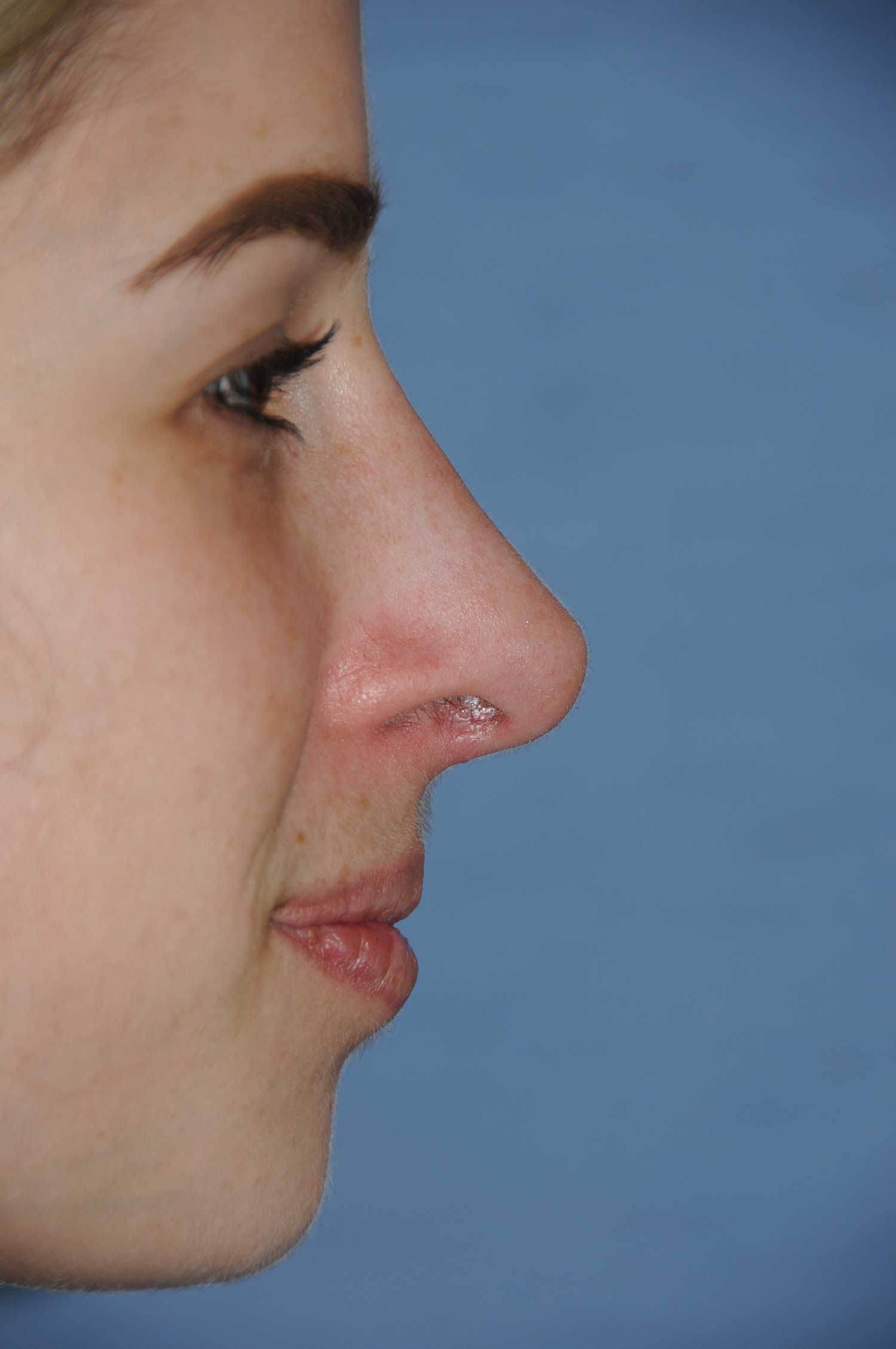
Columella: The tissue between the nostrils at the base of the nose comprising of medial cartilage of the lower lateral cartilages, and the surrounding soft tissue here.
Columellar – Lobular Angle:This is the angle formed by the tissue under the tip, or infratip lobule, and the columella. This ideal angle is about 35-40 degrees from the horizontal.
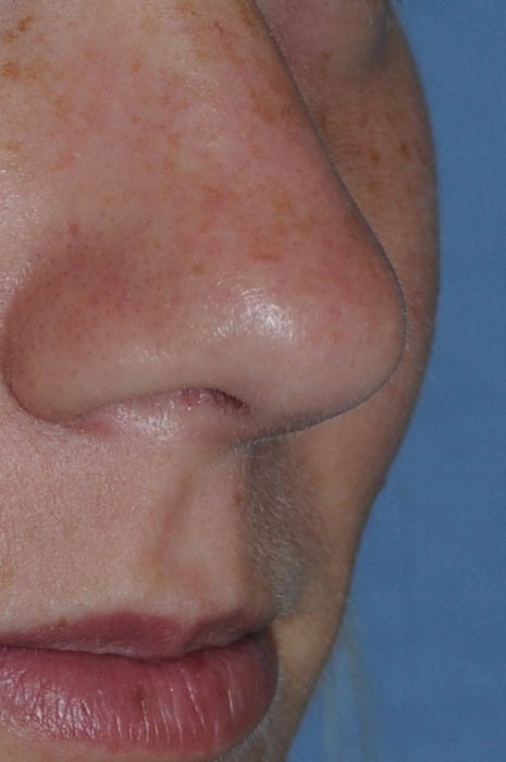
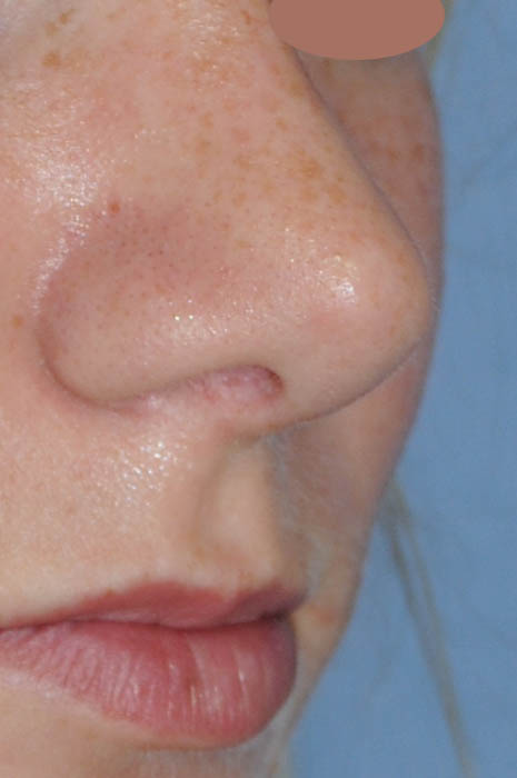
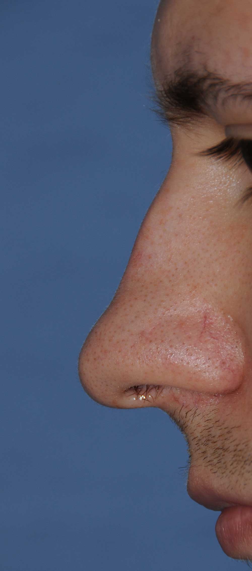
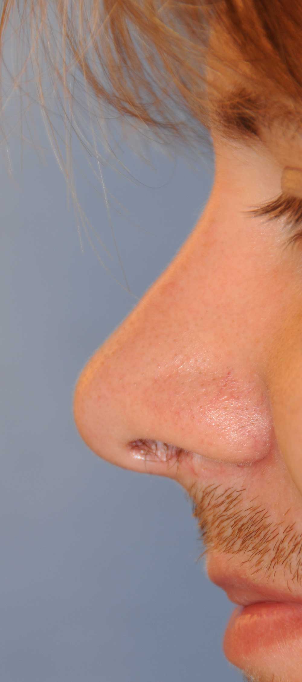
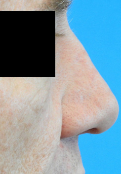
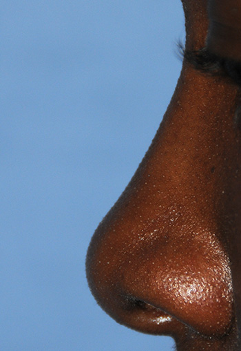
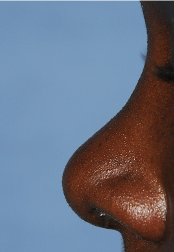
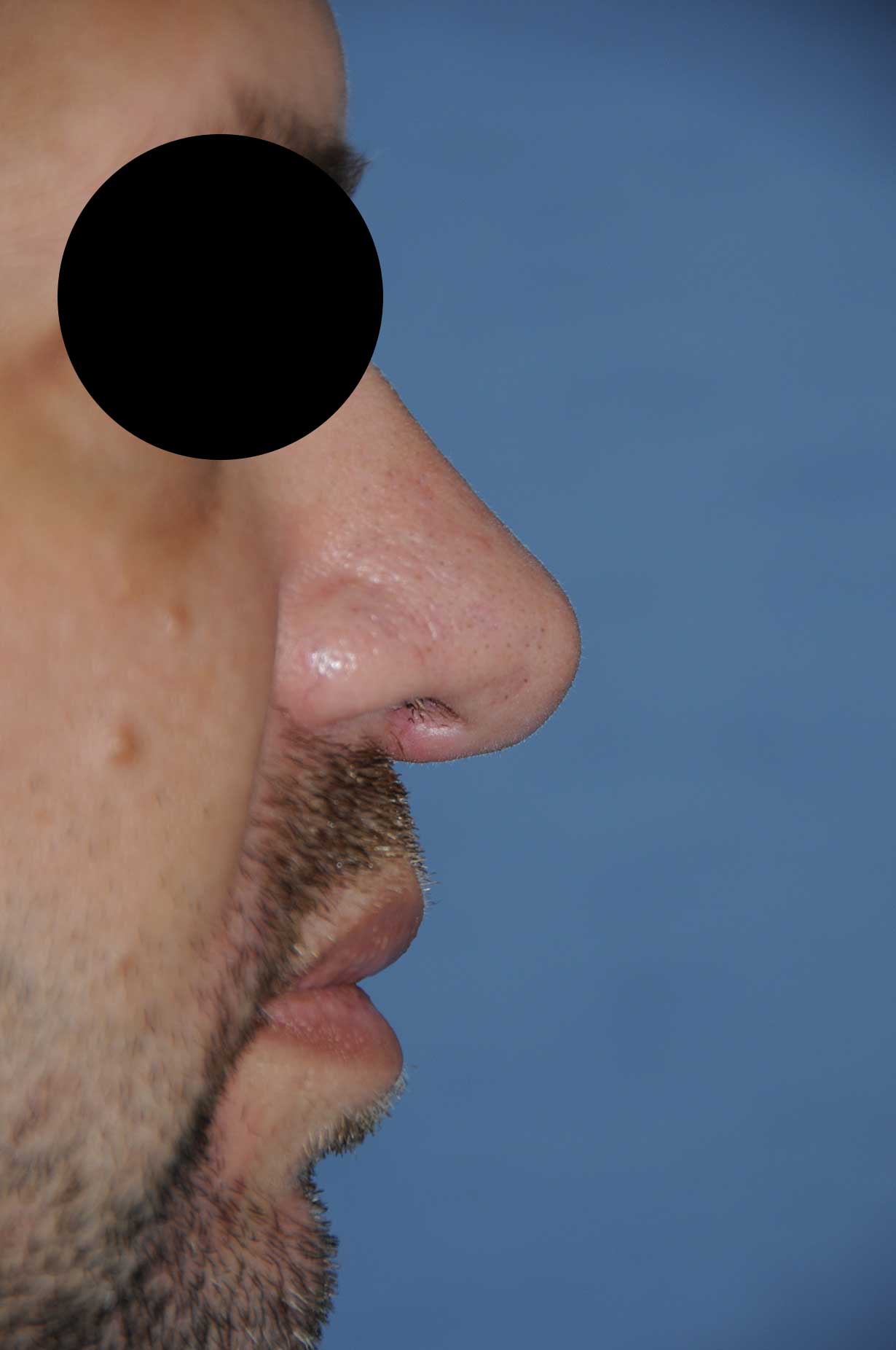
Dorsum: Is the nasal bridge from the glabellar origin to the supratip area. Dorsal, ventral, cephalic and caudal are defined in out Rhinoplasty Anatomy Page
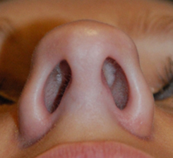
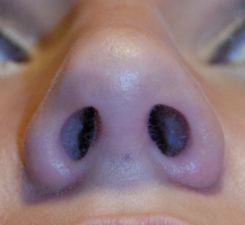
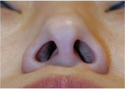
External Nasal Valve: the external opening of the nostril.
Infratip Lobule: The portion of the tip that is between the tip defining points and the columellar lobular junction.
Intercartilaginous Incision: This is an incision that occurs between the upper laterals and lower laterals. This is often used for closed rhinoplasties and for the approach to just the bony bridge and sidewall when adjustments need to be just in this area.
Internal Nasal Valve: This the valve where 50% of all the resistance to breathing occurs. This includes all the breathing including what happens in the lungs. This area is made up of the septum, upper lateral cartilages and the floor of the nose. This is located approximately at the alar groove when you try to locate it from an external landmark. Work in functional nasal breathing and rhinoplasty techniques focus alot of their attention to this area.
Keystone Area: This is the area where the cartilaginous dorsal septum (and the same location of the upper lateral cartilages) connects to the nasal bones.
Limen Vestibuli: This the junction where the caudal edge of the upper laterals meets the lower lateral cartilages. The two cartilaginous sections meet in what is called the scroll area.
Lower Lateral Cartilages: Are the cartilages that are inferior to the upper lateral cartilages. The size, shape, and resilience of the lower lateral cartilages determine the appearance of the tip and lower third of the nose. There are 3 segments to this cartilage structure: the lateral crus, intermediate crus, and the medial crus.
Marginal incision: This is often referred to as the infracartilaginous incision. This incision follows the lower edge of the lower lateral cartilages. This is used with the columellar incision to create the open approach.
Medial Crura: This is the segment of the lateral crura that is at the inferior and caudal end. The connection of the both medial crura and intervening and covering tissue make up the columella.
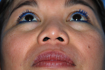
Medial Crura, footplate: This is the segment that extends along the floor of the nostril. When you look from the worms eye view and you look into the nostrils, the medial crura is the soft bend from columella to base of nostril.
Nasal Lobule: This is the area that has the following boundaries the supra tip superiorly, the alae laterally, the anterior edge of the nostril rim and columella posteriorly
Nasion: The soft tissue depression at the junction of the glabella and nasal bridge. This coincides with the Radix which is the bony representation of this point.
Nasolabial angle: This is the angle formed by drawing a line through most anterior point and posterior point of the nostril on lateral view and the vertical plane when the subject is in the frankfort horizontal. In a recent study, the ideal and most aesthetic nasolabial angle ranged from 100.9 to 108.9 degrees in the female nose and 90.7 to 103.3 degrees in the male nose (Plast Reconstr Surg. 2014 Aug;134(2):201-10.The ideal nasolabial angle in rhinoplasty: a preference analysis of the general population. Sinno HH1, Markarian MK, Ibrahim AM, Lin SJ.).
Nostril Base: is the ridge that runs along the floor of the nose at the nostril area from columellar base to alar base. Procedures here involve reducing this area to narrow the nostrils and base as a whole.
Pyriform Aperture: Is the bony window of the nose made up of Maxilla laterally and inferiorly, and nasal bones and nasal process of the frontal bone superiorly. Sometimes opening up the sides of the pyriform aperture will help enlarge the airway for someone with nasal obstruction.
Radix: As noted above, this is the bony element of the nasion. It is located at the most depressed area of the glabellar nasal bone intersection | junction.
Rhinion: Is the area where the nasal bones connect with the cartilaginous elements of the septum and upper lateral cartilages
Rim Incision: is the incision at the edge of the nostrils just inside. Historically, this may be just at the most inferior edge of the nostril. This approach is not as widely used because the closer you get to the edge of the rim the more deformities you are likely to have.
Scroll Area: Is the curl locking of the upper lateral cartilage and lower lateral cartilage. The dynamic relationship allows the internal nasal valve area to have some spring and coil inwards during inspiration and bounce back with expiration.
Sesamoid cartilages: are the variable amounts of cartilage that are located between the upper and lower lateral cartilages laterally.
Soft Tissue Triangle: Is the area between the dome, anterior medial edge of the nostril and the inferior lateral edges of the intermediate and lateral crura | segments.
Subnasale: The junction of the columella and upper lip. This has been a landmark that has been used for facial beauty theories.
Supra Tip Area: This is the area that is superior to the nasal tip. Usually it is more shadowed along with the bridge which give the tip its highlighted prominence.
Tip: The most anterior and forward projection part of the tip area.
Tip, Domes: Part of the Tip that creates two prominent light reflecting points that coincide with the junction of lateral and intermediate crura’s inferior and lateral ends. The domal area creates a diamond shape where the domal area constructs the lateral points and the superior diamond point is the same lateral and intermediate junction at the superior, anterior and medial junction.
Tip, Projection: The distance from the alar base crease from lateral view of the patient to the most anterior projecting part of the nasal tip. The ideal triangle from: 1. Subnasale to the most anterior projecting part of the nasal tip; 2. Length of the nose from Nasion to the Subnasale; 3. Nasion to the most anterior projecting part of the nasal tip should be a 3, 4, 5 ratio.
Tip Rotation: Movement of the Tip in relation to the Nasolabial Angle. An increase in the nasolabial angle indicates more rotation and a smaller angle indicates derotation.
Transfixion Incision: Is an incision made between the medial crura and septal cartilage intervening space. It usually indicates an incision all the way through from nostril to nostril. A hemitransfixion usually indicates an incision on one side without interrupting the tip supporting connection between the septum and medial crura of the lower lateral cartilages.
Upper Lateral Cartilages: These are the paired cartilages that attach to the septum at the nasal bridge in a T shaped relationship. These cartilages make up the middle third. Changing the shape of the upper laterals will affect the middle thirds appearance. These are also vital for the airway at the internal nasal valve, the junction between the septum and the upper laterals most inferior point where the nostrils enter into the nasal cavity.


 Patient Reviews
 Cards From Patients
 Share Your Story